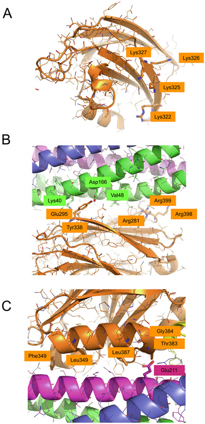
Figure 3. Mutations of the primary and tripartite interfaces.
(A) Close-up view of the polybasic region of Syt1 C2B with residues Lys322, Lys325, Lys326, Lys327 shown as sticks (same view as in the left panel of Figure 2(A)). (B) Close-up view of the primary interface with residues SNAP-25A Lys40, Asp166, Val48 (DEE mutations) and Syt1 C2B Arg281, Glu295, Tyr338, Arg398, Arg399 (quintuple mutant) shown as sticks. (C) Close-up view of the tripartite interface with residues Syt1 C2B Thr383, Gly384, Leu387, Leu394, Phe349 and syntaxin-1A Glu211 shown as sticks. For all illustrations the crystal structure (PDB ID 5W5C) was used.

