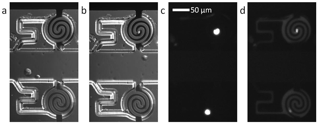
Figure 2.

Brightfield micrographs obtained using differential interference contrast (DIC) of a) two cells captured by DEP and b) the same cells transferred into the adjoining analysis chambers. Fluorescence micrographs of c) the captured cells stained with calcein AM dye and d) the same chambers following application of 148 Vpp lysis voltage. The calcein dye has diffused out of the electroporated cells to illuminate the chambers.
