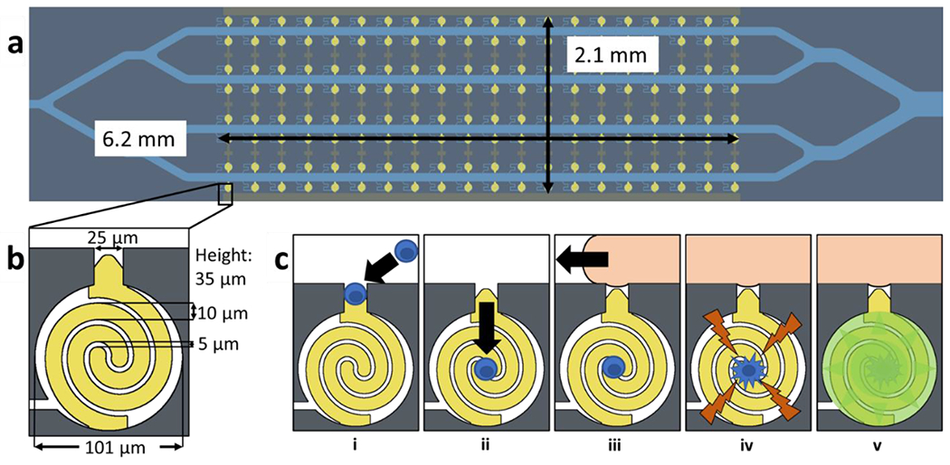
Scheme 1.

a) Illustration of the microfluidic device. The analysis area consists of a 6.2 × 2.1 mm array of 160 chambers b) scheme of electrode and chamber dimensions. Each chamber is a cylinder with a diameter of 101 μm and height of 35 μm. The chambers are connected to the main channels by a 25 × 25 μm capture pocket and a 7 μm wide leak channel. c) scheme of assay process i) DEP cell capture ii) fluidic cell transfer iii) ionic liquid isolation iv) electroporation v) analysis of fluorescence increase.
