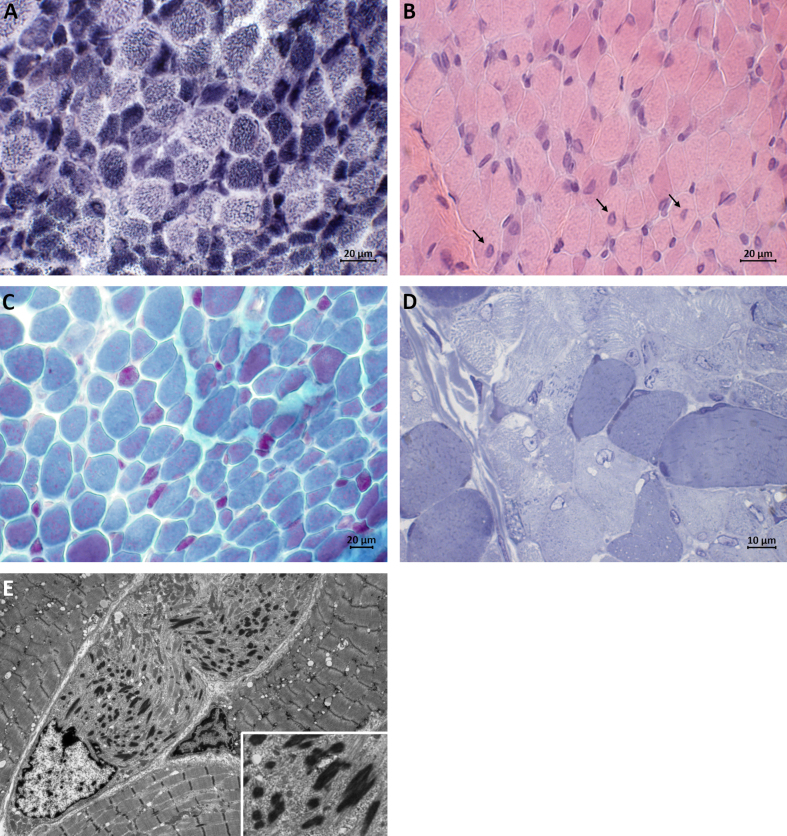
Fig. 1.
Muscle biopsy taken at the age of 7 months. (A) Darkly stained type 1 fibers are predominantly smaller in size than lightly stained type 2 fibers (NADH-TR staining). (B) H&E staining showing central nuclei in several fibers (arrows). (C) Red nemaline bodies are accumulated in small type 1 fibers and scattered rods are seen in larger fibers (Gomori trichrome staining). (D) Semithin section showing darkly staining rod-like structures (Toluidine blue staining). (E) Electron micrograph showing electron-dense ovoid-shaped nemaline rods.

