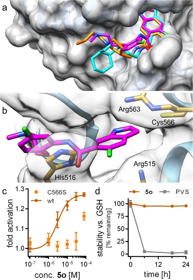
Figure 2.

Predicted binding modes of 5o (magenta), 5r (cyan), and 5v (orange) to the Nurr1 LBD (PDB ID 6dda(21)). (a) The three active DHI descendants 5o, 5r, and 5v were predicted to bind to the DHI-binding site and extend toward a hydrophobic groove lining helix 12. (b) The most active Nurr1 agonist 5o formed a face-to-face contact with His516, which is not observed for 5r and 5v, supporting the higher affinity of 5o. (c) 5o was less active on the Nurr1-C566S mutant, supporting interaction with the proposed epitope. Data are the mean ± S.E.M.; n ≥ 3. (d) 5o was stable against reaction with glutathione (GSH). Phenyl vinyl sulfone (PVS) as positive control (125 μM 5o or PVS were incubated with 2.5 mM GSH in PBS at 37 °C). n = 3.
