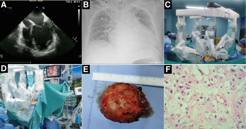
Figure 1.
The main auxiliary examination results and surgical operation diagram of the patient. (A) Echocardiography showed a markedly enlarged left atrium and left ventricle. (B) Chest radiography revealed an enlarged cardiac shadow. (C and D) Da Vinci robot-assisted laparoscopic resection of the adrenal pheochromocytoma. (E) The resected ovoid mass (60 × 60 × 50 mm) with envelope visible by the naked eye. (F) Under a microscope (400 × magnification, hematoxylin-eosin staining), the pathology of the tumor showed cytoplasmic abundance presenting as granules or vacuoles, round or oval nuclei, and visible nucleoli, which are consistent with the features of a pheochromocytoma.

