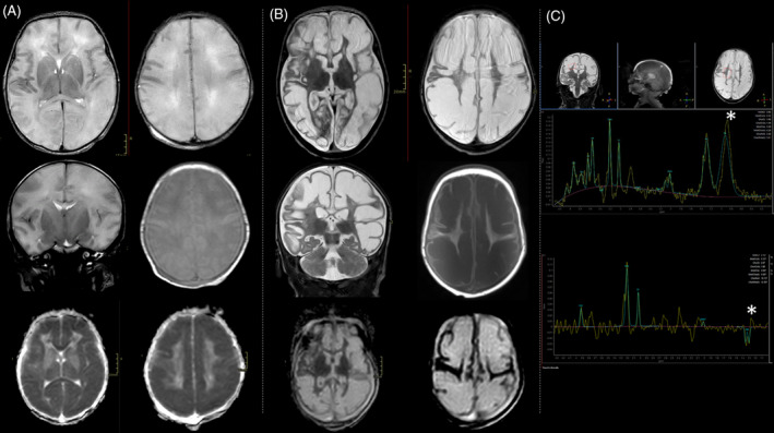
Figure 1.

(A) Brain MRI on day 1 (Transverse and coronal T2 WI, transverse T1 WI, DWI ADC) showed brain swelling with T2 diffuse white matter mild hyperintensity, swollen appearance of the cortical gyri with flattening of the cortical sulci and apparent thinning of the cortex. DWI ADC (bottom) showed significantly restricted diffusivity of the white matter (mean 0,4 mm2 sec). (B) Brain MRI on day 30 (Transverse and coronal T2 WI, transverse T1 WI, DWI ADC) showed evolution toward multicystic encephalomalacia with marked T2 hyperintensity and T1 hypointensity of the white matter. DWI ADC diffusivity considerably increased (3 mm2 sec). (C) MR spectroscopy (short TE‐31 ms– at the top and long TE‐144 ms– at the bottom) exhibited high lactate peak, with typical doublet inverted at long TE (asterisks). There was also an increased peak of choline and reduction of NAA compared to reference values for age.
