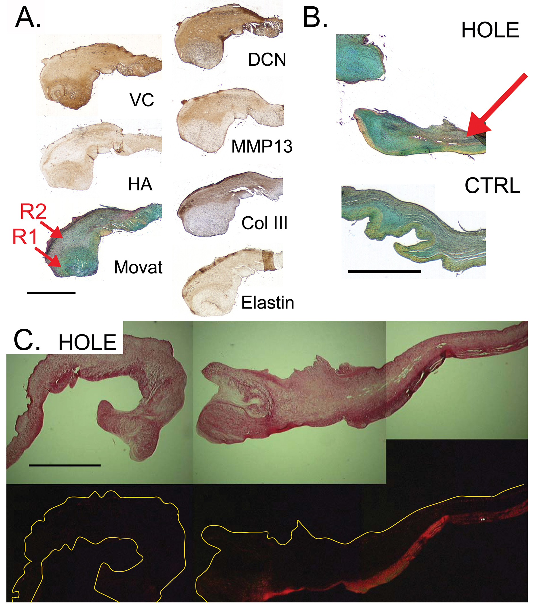
Fig. 5.

A) Two regions of remodeling (R1, R2) were evident as indicated by the red arrows: R1=immediately adjacent to the hole, rich in VC, HA, and demonstrating alcian-blue staining in Movat-stained sections; R2=located interior to R1 relative to the hole-punch, rich in DCN, MMPs, Col III, and elastin. B) Decreased delineation (indicated by red arrow) in HOLE PML compared to CTRL. C) Visualization of picrosirius red-stained tissue under polarized light demonstrates disruption of collagen backbone in HOLE PML. Scale bars for all images represent 1 mm.
