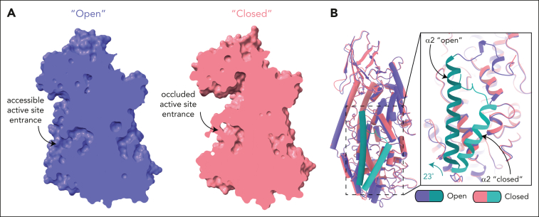
Figure 3.
Conformational changes in the 12-LOX dimer. (A) Surface representation of 12-LOX in the open (left) and closed states (right), showing the active site cavity of the dimer 12-LOX subunits. In the open conformation, the cavity is occupied by a small molecule. (B) An alignment of open and closed states shows a 23° rotation and unwinding of the N-terminal residues of the α2-helix. The inset shows the zoomed-in view of the active site entrance. The α2-helix is in cyan.

