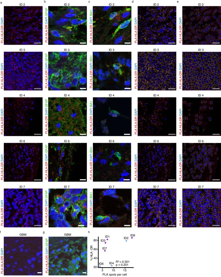
Extended Data Fig. 4. H3K27M neoepitope co-localization with HLA class II-DR on tumor cells and myeloid cells in patients ID 2, ID 3, ID 4, ID 6 and ID 7.
a–c, Proximity ligation assay (PLA) of primary tumor tissue of patient ID 2, ID 3, ID 4, ID6 and ID7 from top to bottom with H3K27M and HLA-DR antibodies (red) in combination with 4’,6-Diamidino-2-phenylindol (DAPI) nuclear staining (blue) alone (a), co-staining with glial fibrillary acidic protein (GFAP) (green) (b) and co-staining with ionized calcium-binding adapter molecule 1 (IBA1) (green) (c). Scale bar in white = 30 μm; in gray = 10 μm. e, f, Automated segmentation of PLA spots (e) and nuclei (f) following rolling ball background subtraction, filtering with gaussian blur and maxima detection of visual field in a. Scale bar in gray = 10 μm. f, g, PLA as in a and co-staining as in b of H3-wildtype glioblastoma. Scale bar in white = 30 μm. h, Pearson correlation of PLA spots per cell with IHC score of HLA-DR expression across 7 patients with available FFPE tissue. Two-sided t-Test was used.

