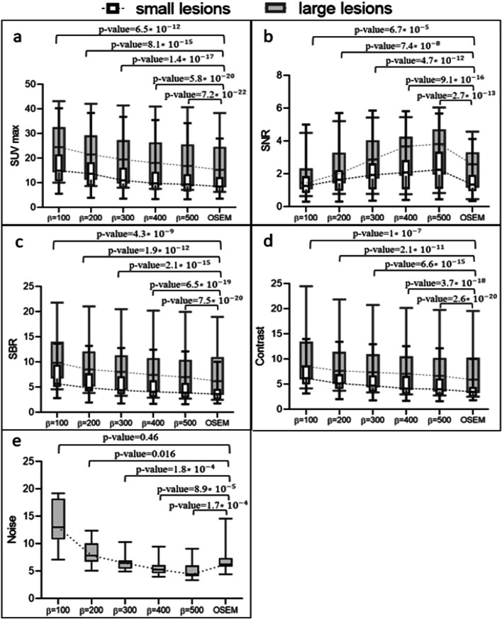
Fig. 6.
a The average SUVmax, b SNR, c SBR, d image contrast, and e noise within the uniform liver area for two categories of lesion sizes (small and large) in a medical research using 18F-FDG. The reconstruction methods used were OSEM and Q.Clear, with β values ranging from 100 to 500 in increments of 100. Dotted lines link the median values. Note that the values depicted in the graphs correspond to the p values derived from the paired t test conducted between the two reconstruction techniques

