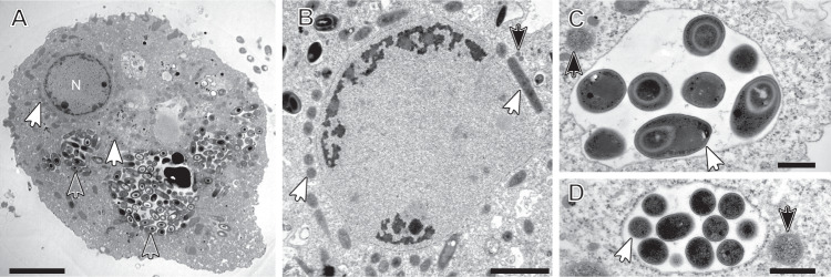
Fig. 1. Transmission electron micrographs of Pelomyxa schiedti cells.
A Overview of P. schiedti cell with the nucleus (N); the different arrowheads indicate different prokaryotic morphotypes; Open arrowheads point to putative Ca. Vesiculincola pelomyxae cells, while white filled arrowheads point to putative Ca. Methanoregula pelomyxae cells. B Detail of the nucleus and the bacterial symbionts located around it; white arrowheads point to putative Ca. Methanoregula pelomyxae cells while black filled arrowheads point to putative MROs. C, D Detail of the bacterial symbionts located in the cytosol (black filled arrowheads) and inside the vacuole (white arrowheads). The bacterial cells in the vacuole (C) are in the process of endospore formation; the diameter of the sporangia is considerable larger than that of the vegetative cells (D) The black filled arrowheads in C and D point to putative Ca. Syntrophus pelomyxae cells. Scale bar: A – 5 µm; B – 2 µm; C, D – 500 nm.

