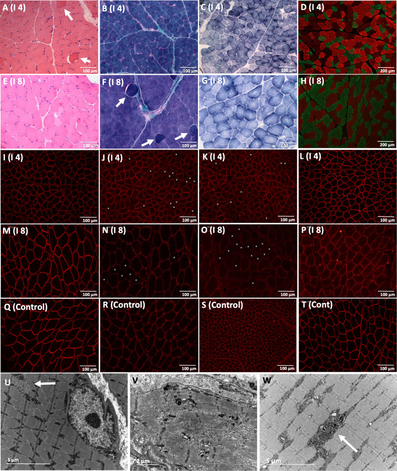
Figure 4.
Myopathic muscle pathology from quadriceps and deltoid biopsies. Biopsy from Individual 4 (A–D, I–L and U–W) was taken from quadriceps at 3 years and biopsy from individual 8 (E–H and M–P) was taken from deltoid at 12 years. Variability in fibre size and fibres with internalised nuclei were observed with H&E stain in individuals 4 and 8 (A, E), as well as hypercontracted fibres (arrow A, F). Increased connective tissue with fibrosis or fatty infiltration were not observed (B, F). Blurred unstained areas in some fibres were noted with SDH (C, G). Selective atrophy of fast fibres is noted with double immunostaining of myosins. Slow-type fibres stained red and fast-type fibres stained green (D, H). Sarcolemmal beta-dystroglycan was normal (I, M) compared with control (Q), which implies that the integrity of the sarcolemma is not altered. A reduction of sarcolemmal alpha-dystroglycan with a mosaic pattern (asterisks in fibres with reduced alpha-dystroglycan) was noted in individuals 4 and 8 with IIH6 (J and N) and VIA 4 (K and O) antibodies, compared with control (R and S). Merosin immunolabelling was normal in individuals 4 (L) and 8 (P) compared with control (T). Electron microscopy (U, V and W) performed in individual 4 showed focal Z-band streamings (arrow in U), a necrotic fibre with a structural disruption of the sarcomere (V) and electron-dense degraded material (arrow in W). Scale bar=100 µm in A–T. Scale bar=5 µm in U and W. Scale bar=2 µm in V.

