Figure 3.
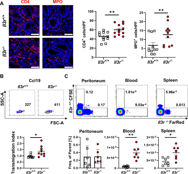
Il3r alters T cell recirculation. (A) Immunofluorescence staining for CD4 (left, red) and MPO (right, red) in colon tissue of Rag1 −/− mice after transfer of naïve CD4+ T cells from Il3r +/+ and Il3r −/− mice counterstained with Hoechst (blue). Left panels: representative images, white arrows highlight CD4+ or MPO+ cells. Right panels: quantification of CD4+ and MPO+ cells per high power field (HPF). n=11–12 per group, unpaired t-test (CD4) and Mann-Whitney (MPO); scale bars – 50 µm. (B) Migration of Il3r −/− or Il3r +/+ thymus T cells over porous membranes towards rm Ccl19. Upper panels: representative flow cytometry. Lower panel: quantification of Ccl19-specific transmigration; n=8–9 per group, Mann-Whitney test. (C) Recirculation of FarRed-stained Il3r −/− and CFSE-stained Il3r +/+ thymus T cells after i.p. injection. Upper panels: representative flow cytometry of peritoneal, blood and splenic cells. Lower panel: quantification; n=6–7 per group, paired t-test. i.p., intraperitoneal; MPO, myeloperoxidase.

