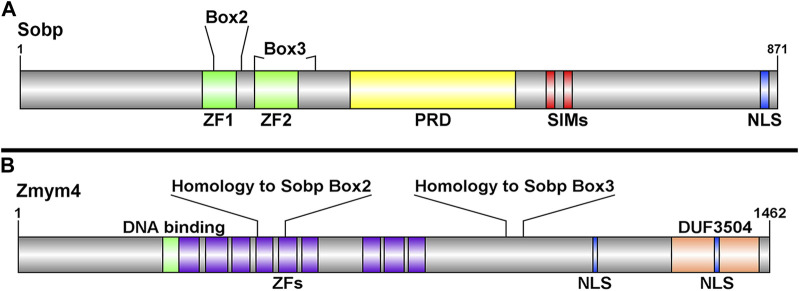
FIGURE 1.
Schematic representations of the Sobp and Zmym4 proteins highlighting their different domains. (A) Sobp protein structure adapted from Tavares et al., 2021 highlighting Box2 and Box3 identified by Kenyon et al., 2005. Sobp contains two zinc fingers (ZF1, ZF2), a proline rich domain (PRD), two SUMO-interacting motifs (SIM), and a functional nuclear localization signal (NLS). (B) The Zmym4 protein contains nine zinc finger MYM-containing domains (purple boxes) identified in human ZMYM4 and aligned by sequence homology (Supplementary Figure S3). The areas of sequence homology with Sobp Box2 and Box3 are indicated. Zmym4 contains two putative NLSs, a glucocorticoid-like DNA binding domain (green box), and a C-terminal DUF3504 domain. The NLSs were identified using cNLS mapper (Kojima and Jurka, 2011), the DNA-binding and DUF3504 domains were identified using InterPro (https://www.ebi.ac.uk/interpro/) and the protein structures were generated using DOG2.0.1 (http://dog.biocuckoo.org/). Numbers on the right of each schematic indicate amino acid length of the protein.

