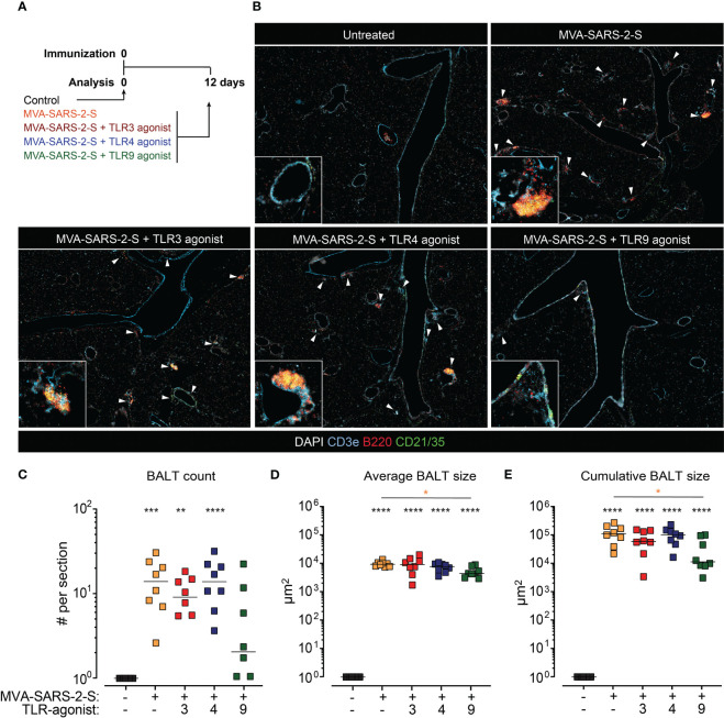
Figure 3.
The addition of TLR9 agonist to MVA-SARS-2-S reduced the formation of bronchus associated lymphoid tissue (BALT). (A) Scheme of immunization protocol. (B) Representative photomicrographs of lung sections reveal induction of BALT (labeled with white arrowheads) 12 days after vaccination. BALT (C) count, (D) average size, and (E) cumulative size calculated as sum of all individual lymphoid structures per lung section. (C–E) Pooled data from four independent experiments with n = 7-8 mice per group. Individual values (symbols) and mean group value (lines). Statistical analysis was done on log-transformed using Brown-Forsythe ANOVA test followed by Dunnett’s T3 multiple comparisons test. *p < 0.05, **p < 0.01, ***p < 0.001, ****p < 0.0001. Black stars - difference to control group; Orange stars – difference between groups receiving vaccine and vaccine mixed with TLR agonists, as denoted by the line.

