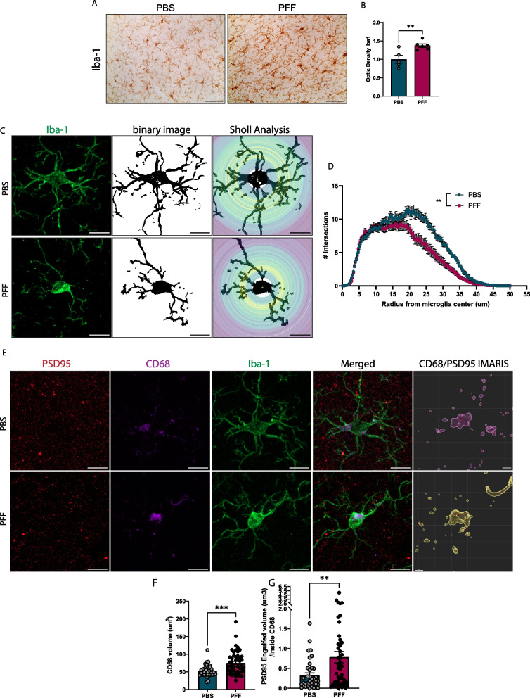
Fig. 4.
Increased microglial synapse phagocytosis. A Increased microgliosis in somatosensory cortex layer 5, detected as Iba1 expression. B Quantification of optical density of Iba1 staining in layer 5. C Representative images of individual microglia for Sholl analysis. Scale bar = 10 µm. D Number of intersections per radius from microglia centers. E 3D reconstruction of PSD95 engulfed inside lysosomal CD68 compartments; scale bar = 10 µm. CD68/PSD95 IMARIS; scale bar = 2 µm. F Quantification of CD68 microglial volume. G Quantification of PSD95 engulfed volume inside CD68. Data in B, D, F, and G are expressed as means ± S.E.M. (n = 37 and 49 microglia for PBS and PFF, respectively, from 5 mice; *p < 0.05, **p < 0.01, ***p < 0.001; Student’s t-test)

