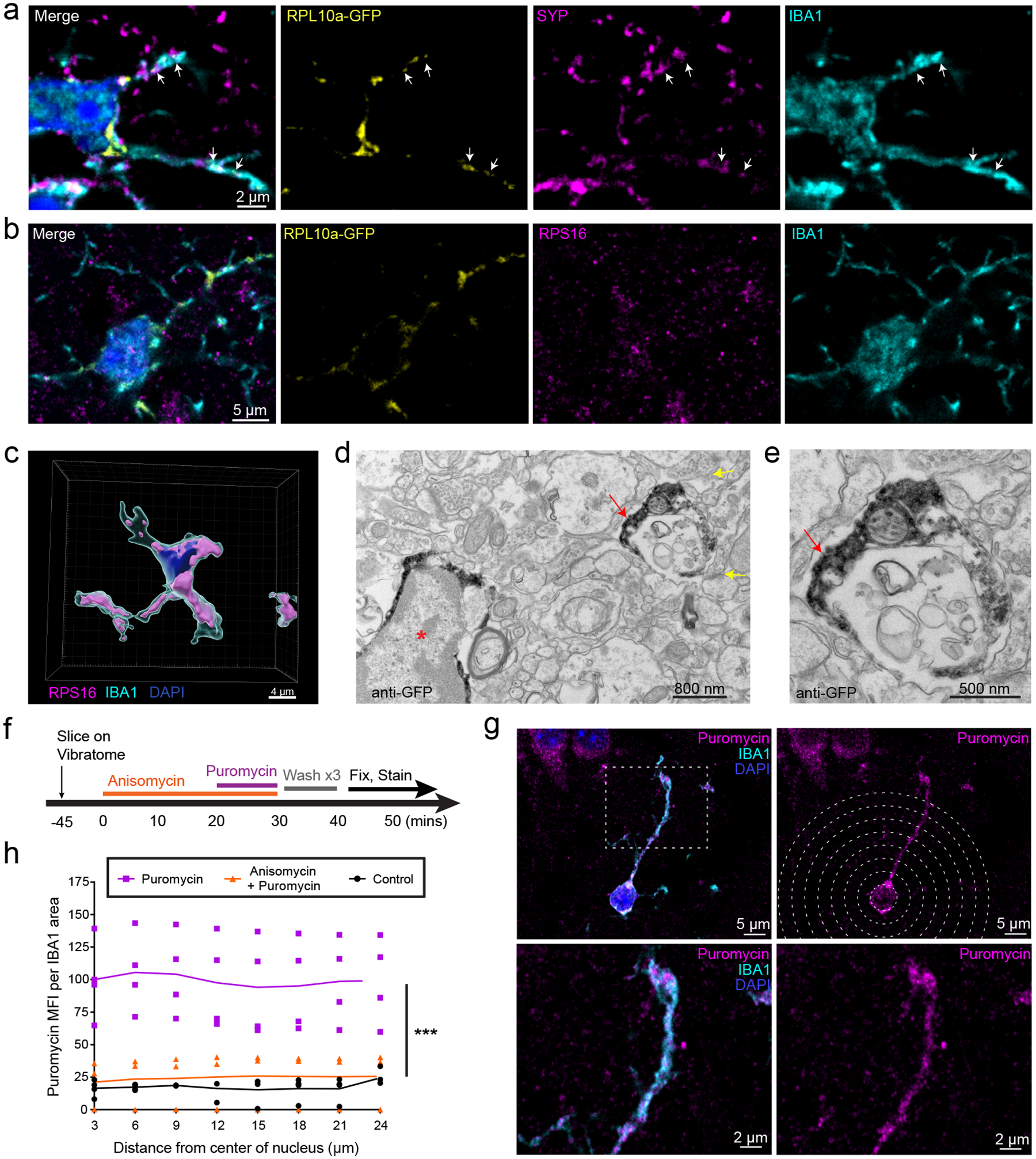
Figure 1: Peripheral Microglial Processes (PeMPs) contain translating ribosomes.

a-e, Brains were harvested from 4–5 week old CX3CR1-Cre(ERT2) x RPL10a-GFP fl/fl mice following tamoxifen induction from p15-p19. a-b, Immunostaining of somatosensory cortex shows localization of ribosomal subunits RPL10a-GFP (a,b) and RPS16 (b) within distal IBA1+ processes and adjacent to synaptophysin positive (a) presynaptic terminals. Images representative of 2 independent experiments from n = 3 mice. c, 3D rendering of RPS16 masked on an IBA1+ cell within somatosensory cortex. Image representative of 1 experiment from n=3 mice. (d,e) Immuno electron micrographs of mouse hippocampus stained with anti-GFP shows RPL10a-GFP present within a microglial soma (red asterisk) and PeMP (red arrow) with nearby synapses (yellow arrows).Images representative of 1 experiment from n = 4 mice. e, PeMP shown at high magnification. f-h, Acute live coronal slices were made from 4–5 week old C57bl6 mice and incubated as shown in the timeline in f. g, Immunostaining for the tRNA analog, puromycin, and IBA1 was performed to quantify local microglial protein synthesis by mean fluorescence intensity of puromycin staining at IBA1-colocalized sites (h), within 3 micron radial bins from the center of the nucleus out to distal PeMPs at increasing distances from the soma (n = 3 anisomycin + puromycin, n = 4 puromycin, n = 4 control brain slices across 2 non-littermate mice with each mouse generating at least 1 slice from each condition). For each slice between 12 and 17 microglia were analyzed and data points represent the mean value of all microglia within the slice while lines represent the means of each treatment group. Treatment is highly significant (p < 0.0001) by two-way ANOVA, while distance (p=0.9996) and interaction of treatment x distance (p=0.9999) are non-significant.
