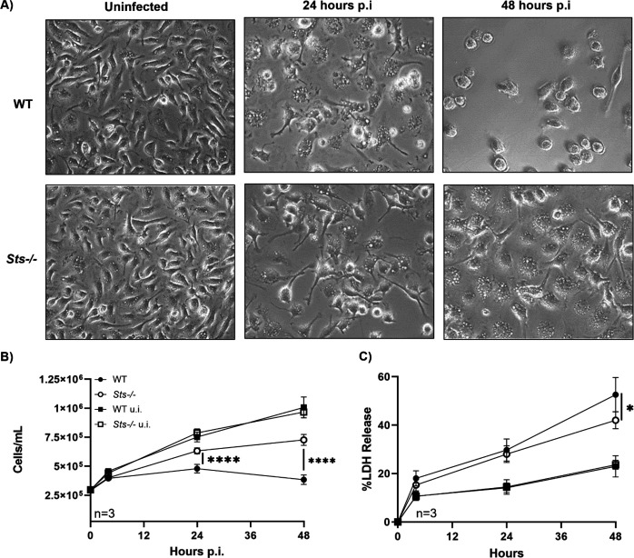
Fig 5.
Reduced S. aureus-induced cytotoxicity exhibited by Sts −/− macrophages. (A) By 48 h p.i., the increased susceptibility of wild-type cells versus Sts −/− to S. aureus-induced cytotoxicity is visible in ex vivo culture. Brightfield images (40×) depict cells before (left) and 24 h (middle) or 48 h (right) post-infection. (B) Uninfected wild-type macrophages (black squares) and Sts −/− macrophages (white squares) display identical growth rates. Infected wild-type macrophages cultures (black circles) exhibit significantly reduced cell numbers relative to infected Sts −/− cultures (white circles). (C) Infected wild-type macrophages release more LDH into culture medium than Sts −/− BMDMs (black and white circles, respectively). LDH release (%) is normalized to LDH in culture medium of lysed BMDMs (100%). (LDH levels in uninfected wild-type and Sts −/− cultures, black and white squares, respectively). Results represent the mean ± SD of three independent experiments. ****, P < 0.0001 by two-way ANOVA and Sidak multiple-comparison test.

