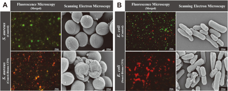
Fig. 6. Fluorescence microscopy and scanning electron microscopy (SEM) images of S. aureus and E. coli in the presence of supernatants.
(A) Microscopic images of S. aureus and (B) E. coli cells. Fluorescence microscopy images present fluorescent-stained S. aureus and E. coli cells after 10 h of cultivation containing 40% CFS of Consti-Biome and Sensi- Biome (Left in A and B). Cells were stained using the LIVE/DEAD Bacterial Viability kit. Live cells (SYTO-9, green) and dead cells (propidium iodide, red). Scale bar indicate 50 μm. SEM images present S. aureus and E. coli cells after 10 h of cultivation (Right in A and B). SEM images show structural damage of S. aureus cultivated in medium containing 40% CFS of Consti- Biome and E. coli in 40% CFS of Sensi-Biome. S. aureus images were observed in the scale of 100 nm with magnification of 100 KX and E. coli images were in the scale of 200 nm with magnification of 50 KX.

