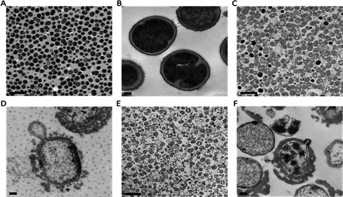
Fig 5.
Transmission electron micrographs of MW2 (MRSA) before and after treatment with exebacase at 8 × MIC (4 µg/mL). Viable cells just prior to the addition of exebacase were viewed at 13,300× (A) and 114,000× (B) magnification (scale bars 2 µm and 100 nm, respectively). Bacteria at 3 min post treatment were viewed at 13,300× (C) and 114,000× (D) magnification (scale bars 2 µm and 100 nm, respectively). Bacteria at 30 min post treatment viewed at 13,300× (E) and 114,000× (F) magnification (scale bars 2 µm and 100 nm, respectively).

