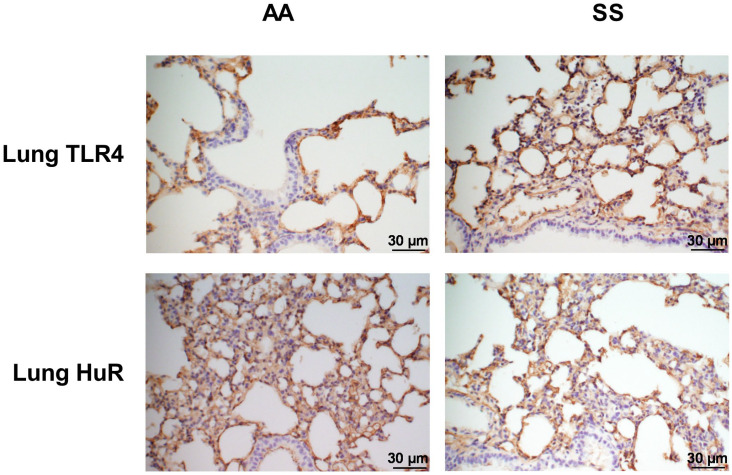
Figure 4.
Immunohistochemical detection of HuR and TLR4 in the lung. Fluorescent (Olympus) microscopy images (magnification ×40) of lung sections stained with an antibody against HuR and TLR4. (A and C) AA mice and (B and D) SS mice. Staining was carried out in different lung sections of untreated 24-week-old mice (n = 4 per group). Scale bars = 30 µm. Blue staining represents nuclei with hematoxylin.

