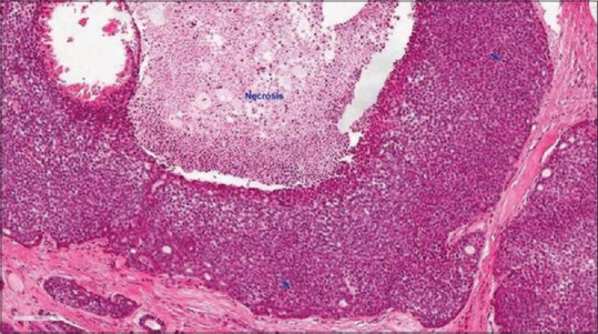
Figure 2.

Histopathological image showing Adcc characterized by tumor necrosis, frequent mitoses (arrows) and marked nuclear atypia with prominent central nucleoli (H&E stain, ×200) (courtesy of pathologyoutlines.com, https://www.pathologyoutlines.com/imgau/salivaryglandsadenoidcysticXu05new.jpg)[35]
