
Figure 3.
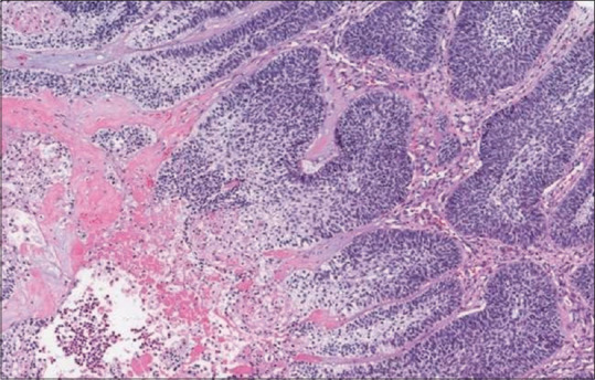
Histopathological image showing ameloblastic carcinoma exhibiting tumor necrosis. (H&E stain, ×200) (courtesy of pathologyoutlines.com, https://www.pathologyoutlines.com/imgau/mandiblemaxillawhoMagliocca1b.jpg)[48]
Official websites use .gov
A
.gov website belongs to an official
government organization in the United States.
Secure .gov websites use HTTPS
A lock (
) or https:// means you've safely
connected to the .gov website. Share sensitive
information only on official, secure websites.
Histopathological image showing ameloblastic carcinoma exhibiting tumor necrosis. (H&E stain, ×200) (courtesy of pathologyoutlines.com, https://www.pathologyoutlines.com/imgau/mandiblemaxillawhoMagliocca1b.jpg)[48]