Fig. 4.
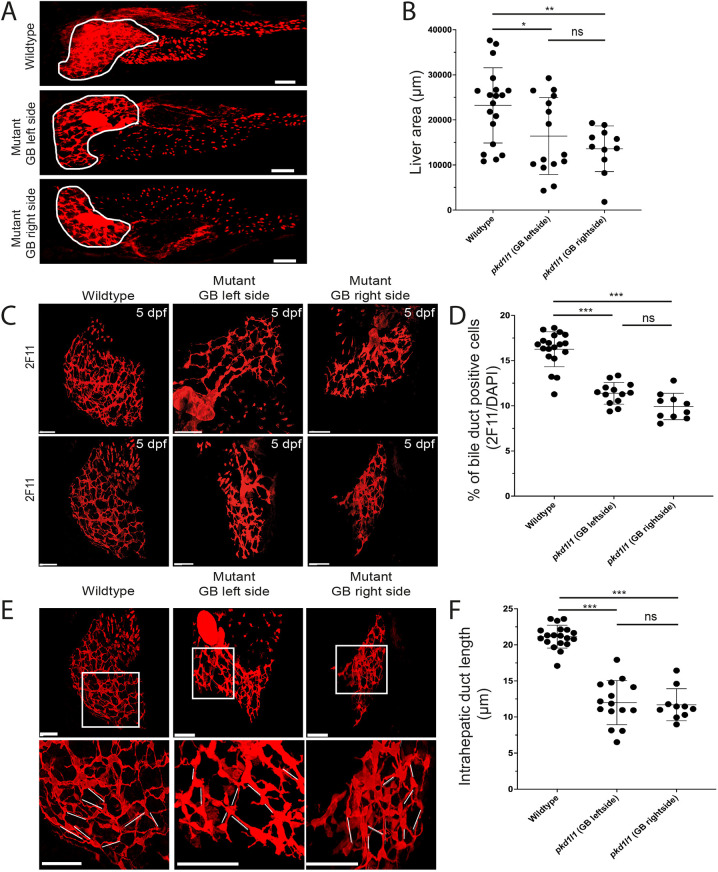
Pkd1l1 function is necessary for normal bile duct development. (A) Confocal images showing bile ducts (assessed by 2F11 antibody staining) in liver (white outline) in wild type and mutants. GB, gallbladder. (B) Quantification showed reduction of the area of the liver in mutants compared to that in the wild type. (C,D) Whole-mount immunostaining of zebrafish livers (C) showed a decrease number of biliary epithelial cells (BECs) and abnormal intrahepatic biliary network in pkd1l1 mutant larvae compared to those in wild-type larvae (D). The numbers of BECs were measured by counting 2F11 and DAPI double-positive cells and the percentages were calculated by dividing by the total number of DAPI-positive cells from each fish. (E,F) Confocal images (E) showed a reduced length of intrahepatic ducts (white lines) in mutants compared to that in the wild type. The lengths of intrahepatic ducts were calculated by measuring the duct distance between two cell bodies and the average of ten ducts was calculated per image (fish) (F). Graphs show the mean±s.d. Scale bars: 50 µm. ns, not significant; *P<0.05; **P<0.01; ***P<0.001 (one-way ANOVA with Tukey post hoc test).

