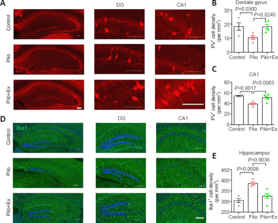
Figure 2.

Exercise suppresses neuroinflammation and restores PV-interneurons in the mouse model of epilepsy.
(A) Immunofluorescence images of PV-interneurons (mCherry, red) in hippocampal DG and CA1. (B) PV-interneuron density in the DG was lower in the Pilo group than in controls, and higher in the Pilo+Ex group than in the Pilo group (one-way analysis of variance followed by Tukey’s post hoc comparisons, F(2,11) = 6.365, P = 0.0146). (C) PV-interneurons in the CA1 were also preserved after treadmill exercise (one-way ANOVA followed by Tukey’s post hoc comparisons; F(2,11) = 12.96, P = 0.0013). (D) Immunofluorescence staining of microglial cells by Iba-1 (green, Alexa Fluor® 594). Scale bars: 100 μm in A and D. (E) Microglial population was larger than normal after pilocarpine treatment and this effect was suppressed after exercise (one-way ANOVA followed by Tukey’s post hoc comparisons; F(2,11) = 14.93, P = 0.0007). All data are presented as mean ± SEM (n = 4, 5, and 5 mice in control, Pilo, and Pilo+Ex groups, respectively). ANOVA: Analysis of variance; DG: dentate gyrus; Ex: exercise; Iba-1: ionized calcium-binding adapter molecule 1; Pilo: pilocarpine; PV: parvalbumin.
