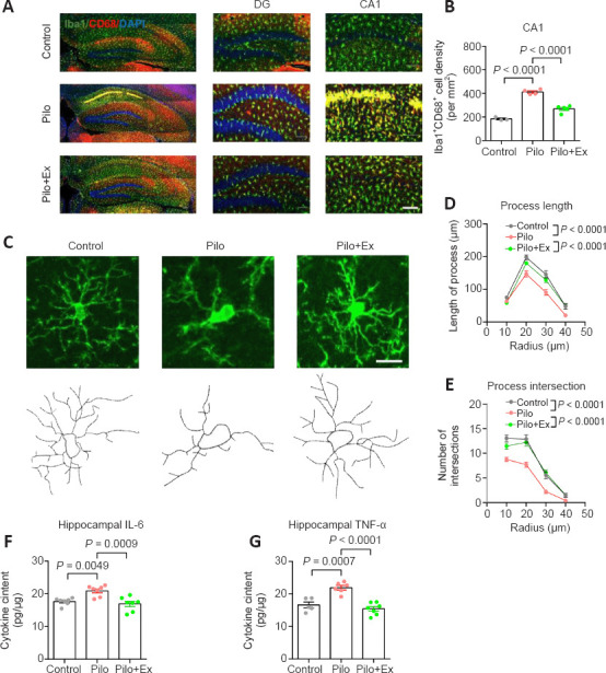
Figure 3.

Activation of microglial cells and neuroinflammation in the mouse model of epilepsy with regular exercise.
(A) Immunofluorescence staining of microglial cells and their reactivity via Iba-1 (GFP, green) co-labeling with CD68 (mCherry, red). Scale bar: 100 μm. (B) The activated microglial population was significantly suppressed after regular exercise (one-way ANOVA followed by Tukey’s post hoc comparisons; F(2,9) = 87.29, P < 0.0001. n = 3, 4 and 5 mice in control, Pilo, and Pilo+Ex groups, respectively). (C) Upper panels, fluorescent images of representative microglial cells. Lower panels, reconstructions of cellular process contours. Scale bar: 20 μm. (D) The total microglia process length was smaller than normal in neurons from the Pilo group, and this effect was absent in neurons from the Pilo+Ex group (two-way ANOVA with respect to the interaction between group and radius factor; F(6,343) = 2.923, P = 0.0086). (E) Microglial process intersected fewer arbitrary concentric circles in neurons from the Pilo group, and those effects were absent from the Pilo+Ex group (two-way ANOVA with respect to the interaction effect; F(6,343) = 3.230, P = 0.0042). N = 20 cells from four mice in each group in D and E. (F) IL-6 concentration in the hippocampus was elevated in the Pilo group and not elevated in the Pilo+Ex group (one-way ANOVA followed by Tukey’s post hoc comparisons; F(2,18) = 11.24, P = 0.0007). (G) TNF-α levels in the Pilo+Ex groups were also lower than those in the Pilo group (one-way ANOVA followed by Tukey’s post hoc comparisons, F(2,18) = 21.96, P < 0.0001). n = 7 mice per group in F and G. All data are presented as mean ± SEM. ANOVA: Analysis of variance; DG: dentate gyrus; Ex: exercise; GFP: green fluorescent protein; Iba-1: ionized calcium-binding adapter molecule 1; IL-6 interleukin-6; Pilo: pilocarpine; TNF-α: tumor necrosis factor-α.
