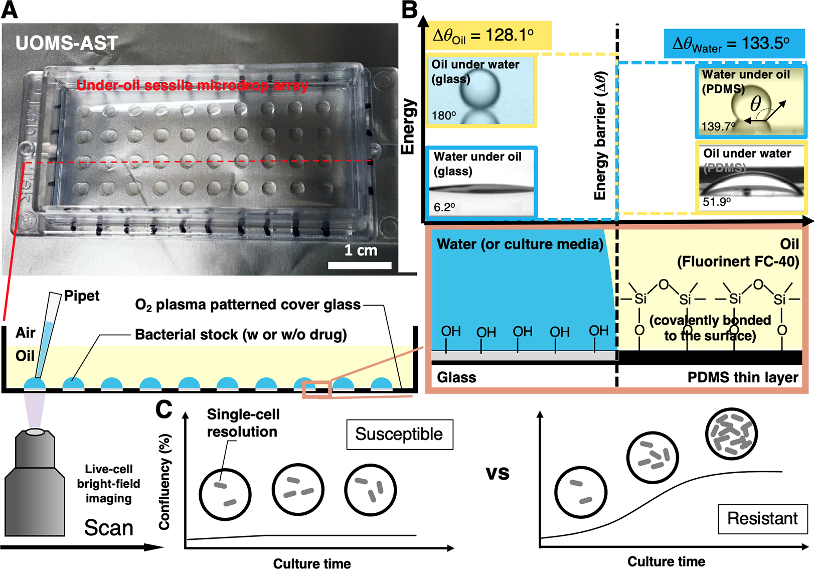
Fig. 1.

The configuration and physics of UOMS-AST. (A) A UOMS-AST device fabricated with a 1-well chambered coverglass (Nunc Lab-Tek - borosilicate glass #1.0 - medical grade silicone adhesive, 6 cm × 2.4 cm, 0.13–0.17 mm in bottom thickness). The glass surface was patterned for an array (4 × 10) of underoil (Fluorinert FC-40, or FC40, 1.5 mL per 1-well chambered coverglass) sessile (i.e., surface-attached) microdrops (2 mm in diameter). 2 μL of bacterial stock with or without antimicrobial was inoculated to each spot under oil by regular single-channel pipetting. (B) A schematic shows the surface chemistry contrast for liquid confinement with the energy (or virtual) barrier. The glass surface was modified by a monolayer of covalently bonded, polydimethylsiloxane (PDMS)-silane molecules and then selectively patterned by oxygen plasma (Fig. S1). (C) A schematic shows the live-cell, bright-field imaging in UOMS-AST and the detection of antimicrobial susceptibility/resistance based on confluency of bacterial cells in the field of view.
