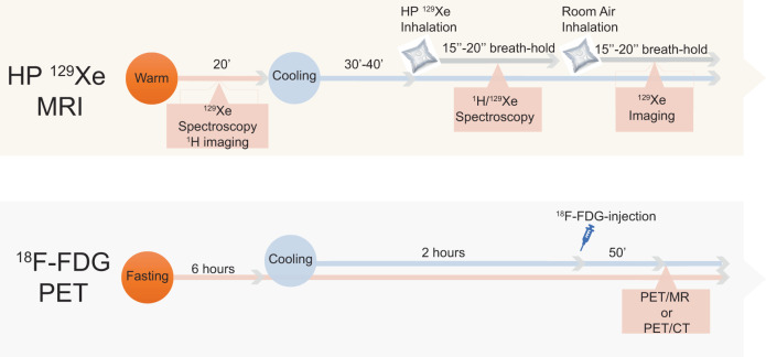
Fig. 2. Flow chart of the human study protocol used in this study.
For the hyperpolarized xenon (HPXe) MRI procedure, participants were exposed to cold for 30–40 min before 1H and 129Xe spectroscopy studies were performed. In this case, water temperature was set at 18 °C and lowered until they started to shiver. When that occurred, temperature was raised until shivering subsided. Three participants were first exposed to warm condition during which a baseline 129Xe spectroscopy study was performed before cold exposure began. For the 18FDG-PET study, participants were asked to fast for at least 6 h prior to arriving at the imaging center. After changing into scrubs, they were wrapped with water perfused cooling pads (same as the ones used for the MRI study). Initial water temperature setting was 18 °C. Temperature was then lowered every 5 min until they began to shiver and then raised by 1 °C every 5 min until shivering stopped. In all cases, all participants were exposed to cold temperatures (≤18 °C) for at least 2 h before 18F-FDG injection and during the PET scan.

