
FIGURE 1.
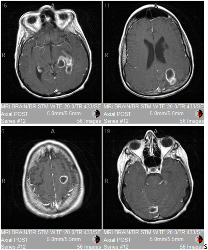
Brain MRI demonstrated intra‐axial ring‐enhancing lesions in the left posterior frontal lobe, left posterior parietal lobe, right occipital lobe and left temporal lobe, all with restricted diffusion and surrounding edema.
Official websites use .gov
A
.gov website belongs to an official
government organization in the United States.
Secure .gov websites use HTTPS
A lock (
) or https:// means you've safely
connected to the .gov website. Share sensitive
information only on official, secure websites.
Brain MRI demonstrated intra‐axial ring‐enhancing lesions in the left posterior frontal lobe, left posterior parietal lobe, right occipital lobe and left temporal lobe, all with restricted diffusion and surrounding edema.