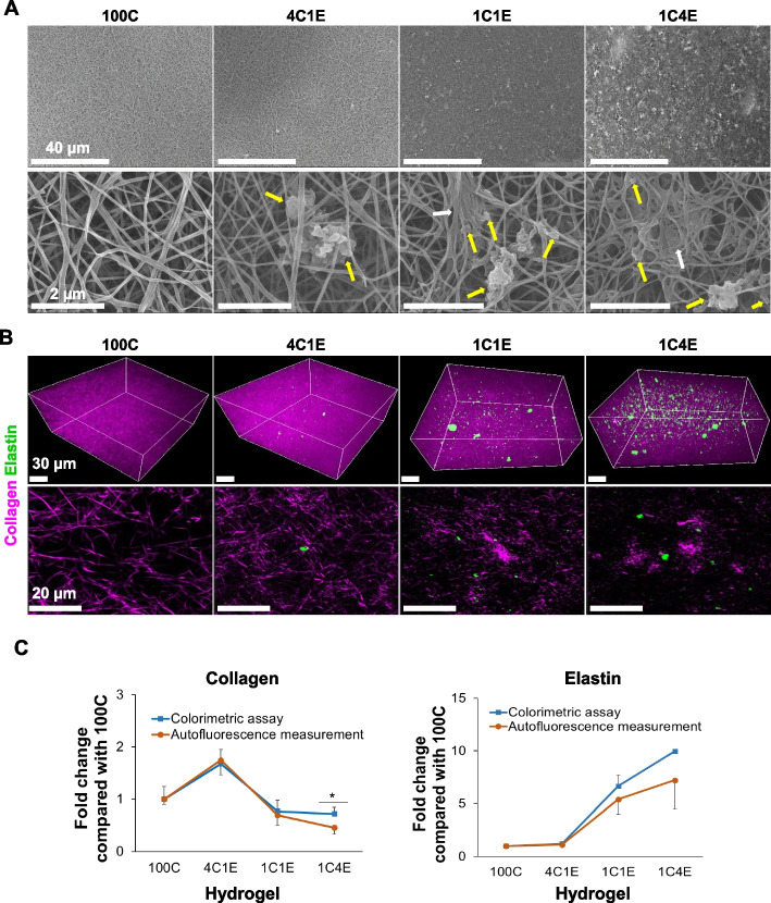
Fig. 1.
Morphology of elastin-variable hydrogels without cells at 0 h. A Scanning electron microscope (SEM) imaging of hydrogels without cells. The yellow arrows indicate insoluble elastin, and the white arrows point to clumps of collagen. B Multimodal nonlinear optical (MNLO) imaging of hydrogels without cells. The purple second harmonic generation (SHG) signal represents collagen, and the green two-photon excitation fluorescence (TPEF) signal represents elastin. C Levels of collagen and elastin in hydrogels at 0 h, as measured by colorimetric assays and autofluorescence intensity analysis. Data are represented as mean ± standard deviation (SD) of triplicate analyses. * p < 0.05

