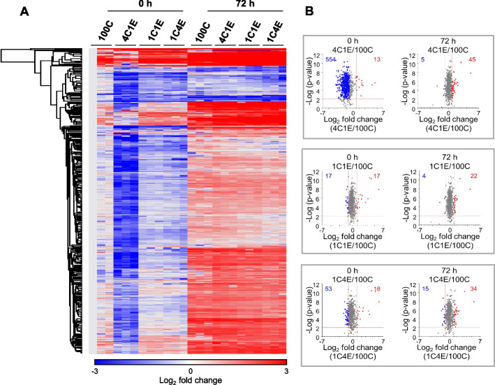
Fig. 3.
Label-free quantification-based proteomic analyses of HDFs in four elastin-variable hydrogels at early and late stages by nLC-ESI–MS/MS. A Heat map of the differentially expressed proteins with at least a ± 1.5-fold change. B Volcano plots of HDFs in elastin-variable hydrogels compared with control (100C). The results show the proteins log2 fold change plotted against the –log10 p-value. Blue and red colors in the heat map and volcano plots represent downregulated and upregulated proteins in the elastin-variable hydrogels compared to 100C, respectively. Red dotted lines indicate p = 0.01 and ± 1.5-fold change

