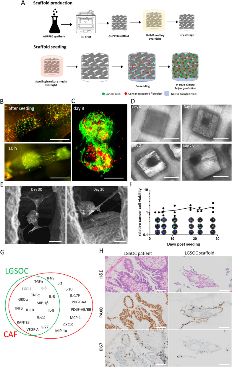
Fig. 2.
LGSOC scaffold model. A Schematic overview of scaffold production and seeding procedure, single cell mixture of cancer cells and CAF suspended in type I collagen solution are added to the scaffold by drip seeding. During in vitro culture cells self-assemble into spheroids, CAF (red) organize at the center surrounded by cancer cells (green). B Fluorescent images of PM-LGSOC-01 Luc eGFP (green) and CAF (red) seeded on a AUPPEG8K scaffold, immediately and 10 h after seeding. Round cells (after seeding) become elongated, indication of migration along the collagen fibers, and self-assembly into a spheroid (10 h). Scale bar are 100 µm. C Confocal image of the LGSOC scaffold model 1 week post seeding on a AUPPEG8K scaffold. Two spheroids within one pore are visualized; red labeled CAF form the center and LGSOC (green) surround the CAF. Scale bare are 50 µm. D Phase contrast images of the LGSOC scaffold model at day 0, 1, 2 and 25 post seeding. Scale bars are 200 µm. E SEM images of the LGSOC scaffold model 1 month post seeding. F Relative cancer cell viability within the LGSOC scaffold model determined by bioluminescence imaging (BLI). G Venn diagram of cytokines identified by Luminex in the secretome of the LGSOC scaffold model. Cytokines are produced by LGSOC cells and/or CAF. H H&E, PAX8 (ovarian cancer marker) and Ki67 (proliferation marker) staining of an LGSOC patient sample and the LGSOC scaffold model (> day 30). Note that the early-passage LGSOC cell culture used in the scaffold is established from the patient sample used to evaluate morphology and PAX8 and Ki67 positivity. Scalebar 100 µm

