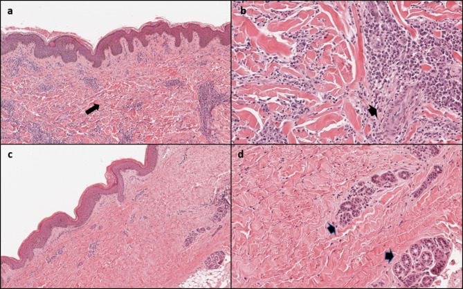
Fig. 2.
Comparison of histological characteristics of juvenile localized scleroderma and connective tissue nevus. Juvenile localized scleroderma: (a) compact fibrosis involving dermal and subcutaneous layers (long arrow), disappearance of skin adnexa, (b) perivascular inflammatory infiltrates (short arrow). Non-familial collagenoma: (c) thickened collagen bundles arranged randomly in the reticular dermis (long arrow), (d) preserved skin adnexa and absence of inflammatory infiltrate are evident (short arrow) (Hematoxylin Eosin, original magnification a-c: 70x, b-d: 200x)

