Figure 1.

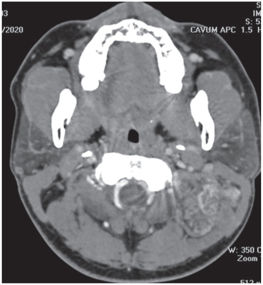
Axial CT contrast scan showed a, well-defined mass with moderate and heterogeneous enhancement in the left semispinalis without calcifications.

Axial CT contrast scan showed a, well-defined mass with moderate and heterogeneous enhancement in the left semispinalis without calcifications.