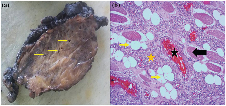
Figure 3.
Intramuscular hemangioma. (a) Grossly, the tumor has a heterogenous aspect with purple hemorrhagic areas, yellowish areas, and medium-sized vessels (yellow arrows). (b) Microscopically, the tumor shows skeletal muscle (black arrow) infiltrated by vascular channels, often small sized, (yellow asterisk), sometimes larger (black asterisk), filled with red blood cells. Note the presence of entrapped fat tissue (yellow arrow) (HE × 100).

