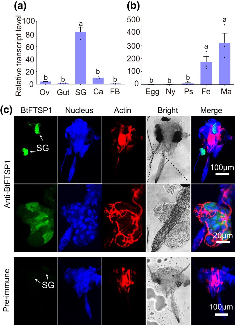
Fig. 3.
Expression patterns of BtFTSP1. a) qRT-PCR suggested that BtFTSP1 was mainly expressed in the salivary gland. SG, salivary gland; Ca, carcass; FB, fat body; Ov, ovary. b) Expression patterns of BtFTSP1 at different developmental stage. Ny, nymph; Ps, pseudo chrysalis; Fe, female adult; Ma, male adult. c) Immunohistochemical staining of BtFTSP1 in B. tabaci head. The insect heads were incubated with anti-BtFTSP1 serum or pre-immune serum conjugated with Alexa Fluor 488 NHS Ester (green) and actin dye phalloidin rhodamine (red) and examined by Leica SP8. The nucleus was stained with DAPI (blue). The lower images represent the enlarged images of the boxed area in the upper image. The boxed area was indicated in a bright filed image.

