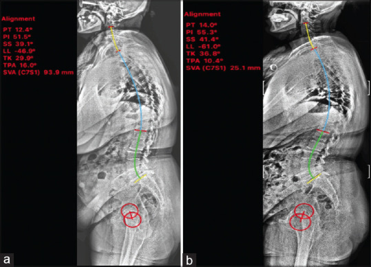
Figure 1.

Preoperative and postoperative spinopelvic parameters of one patient measured via Surgimap program. whole spine radiograph before surgery (a) and after surgery (b), red circles represent femoral heads. There was an increase in lumbar lordosis, increase in thoracic kyphosis, and decrease in sagittal vertical axis values. PT - Pelvic tilt, PI - Pelvic incidence, SS - Sacral slope, LL - Lumbar lordosis, TK - Thoracic kyphosis, TPA - T1-pelvic angle, SVA - Sagittal vertical axis
