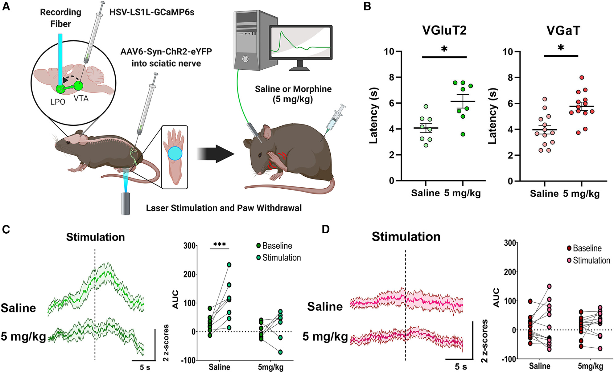
Figure 1. Inputs from LPO VGluT2 neurons to the VTA signal nociceptive stimuli.

(A) Diagram of surgical procedures and recording task. Retrograde Cre-dependent HSV-LS1L-GCaMP6s viral vector was injected into the VTA of vglut2:cre or vgat:cre mice and retrograde AAV6-Syn-ChR2-eYFP viral vector was injected into the sciatic nerve. An optic fiber was implanted over the LPO for fiber photometry calcium imaging recordings of LPO-VGluT2 or LPO-VGaT neurons innervating the VTA in response to laser-induced nocifensive responses after injection of saline or morphine (5 mg/kg).
(B) Latency to paw withdrawal induced by sciatic nerve laser stimulation in vglut2:cre or vgat:cre mice after injections of saline or morphine (5 mg/kg) (mixed ANOVA; VGluT2 n = 8, F(2,23) = 8.516, p = 0.007; VGaT n = 13, F(2,38) = 6.61, p = 0.005; *p < 0.05; data represent the mean ± SEM).
(C) LPO-VGluT2 neurons innervating the VTA signal the nociceptive stimulation of the paw, which is blocked by morphine (5 mg/kg) (***p < 0.001).
(D) LPO-VGaT neurons innervating the VTA do not show responses to the nociceptive stimulation of the paw or morphine (mixed ANOVA; F(2,38) = 3.123, p = 0.032; data represent the mean ± SEM). (See also Figures S2 and S3.)
