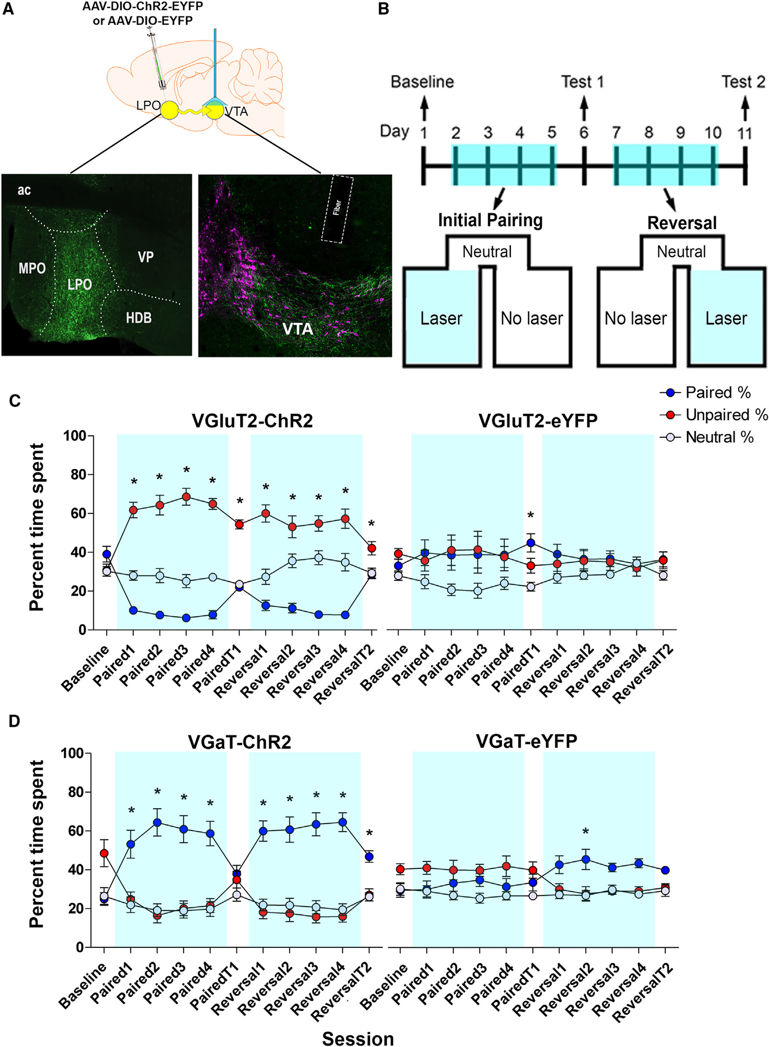
Figure 3. VTA photostimulation of LPO-VGaT fibers is rewarding and VTA photostimulation of LPO-VGluT2 fibers is aversive.

(A) Diagram of virus injection (AAV5-EF1αa-DIO-ChR2-eYFP or AAV5-EF1α-DIO-eYFP) into the LPO and VTA photostimulation of LPO-VGluT2 or LPO-VGaT (top). Histological verification of the virus injection within the LPO and optical fiber placements within the VTA is shown. Boundaries of the VTA are shown by tyrosine hydroxylase detection (TH; magenta) and eYFP shows LPO axons (green) (bottom).
(B) Timeline for the place conditioning procedure. Mice were tested for baseline preference, followed by four pairing sessions (Paired 1–4) and a stimulation-free test (Paired T1). Subsequently, the paired compartment was reversed for four reversal sessions (Reversal 1–4) and a stimulation-free test (Reversal T2).
(C) VGluT2-ChR2 mice with VTA photostimulation of LPO-VGluT2 fibers spent less time in the chamber paired with photostimulation on conditioning days (blue line) and exhibited place aversion during stimulation-free test sessions (n = 14 mice). VGuT2-eYFP-mice with VTA photostimulation of LPO glutamate fibers spent equal times in the photostimulation paired and unpaired chambers (n = 7 mice) (generalized linear model; n = 21; chamber × session × group interaction, χ2(20) = 105.992, p = 1.0503E—13; *p < 0.05; data represent the mean ± SEM).
(D) VGaT-ChR2 mice with VTA photostimulation of LPO-VGaT fibers spent more time in the photostimulation-paired chamber during conditioning (blue line) sessions without developing conditioned place preference for the photostimulation-paired chamber (n = 14 mice). VGaT-eYFP mice with VTA photostimulation of LPO-VGaT fibers did not show a preference for either the paired or the unpaired chamber (n = 7 mice). Data are shown as the mean ± SEM (generalized linear model; n = 21; chamber × session × group interaction, χ2(20) = 42.28, p = 0.003; *p < 0.05). (See also Figure S2.) LPO, lateral preoptic area; MPO, medial preoptic area; VP, ventral pallidum; HDB, horizontal diagonal band; ac, anterior commissure; VTA, ventral tegmental area.
