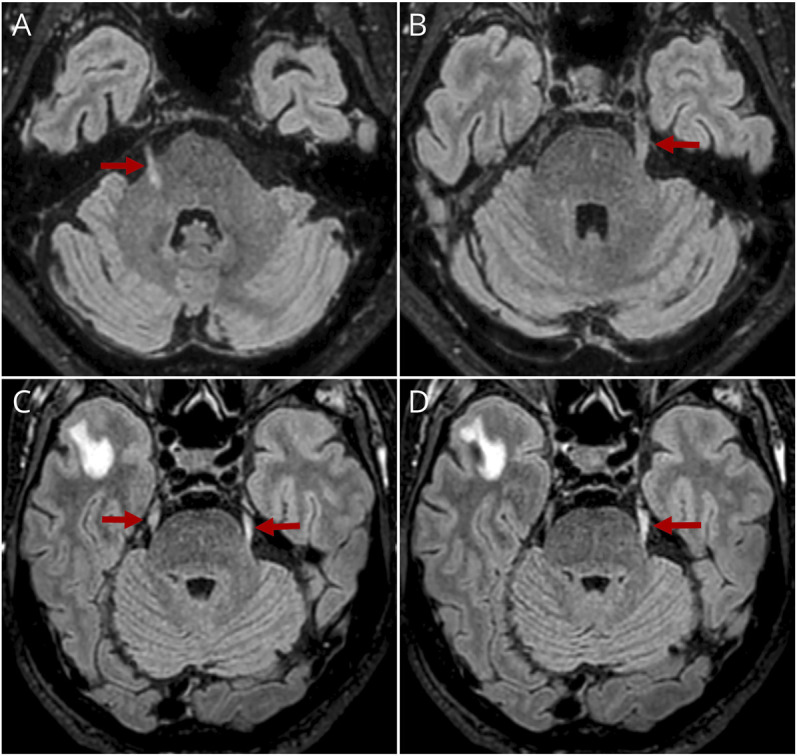
Figure 1. Trigeminal Nerve Abnormalities on MRI of the Brain.

Axial T2 FLAIR sequences showing hyperintensity and slight nerve enlargement of both trigeminal nerves in patients 1 (A, B) and 2 (C, D) (red arrows), including the intra-axial portion of the right trigeminal nerve in patient 1 (A). In patient 2, the right temporal pole abnormalities were attributed to coincidental opercular enlargement of perivascular space with peripheral gliosis.
