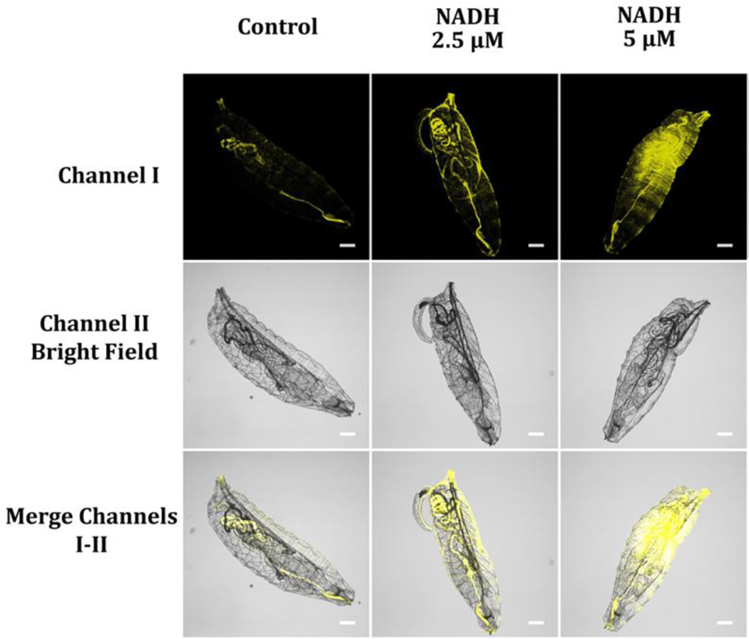
Figure 15.
Freshly starvation-hatched fruit fly larvae were placed in different NADH concentrations from 0 (control) to 5 μM in a pH 7.4 PBS buffer for 15 min, washed three times with the PBS buffer, and then immersed in PBS buffer solution holding 10 μM probe A for 30 minutes. Fluorescence signals from the images were obtained at a wavelength range of 550–650 nm upon 488 nm excitation.

