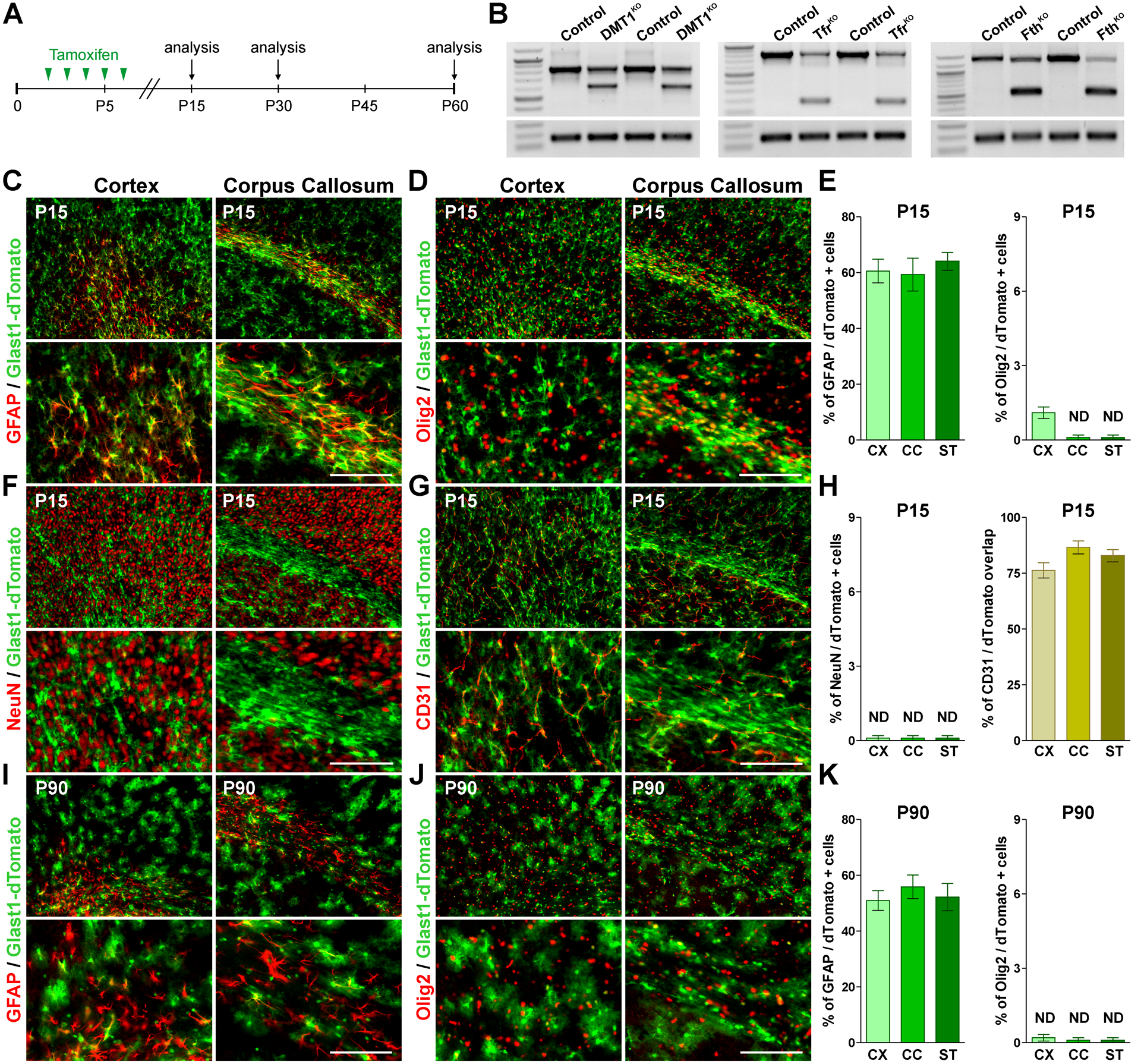
FIGURE 1: Recombination efficiency in the postnatal Glast1-dTomato mouse.

(A) P2 Glast1-KO lines and control (Cre-negative) littermates received 5 consecutive tamoxifen injections and brain tissue was collected at P15, P30 and P60. (B) Semi-quantitative RT-PCRs for DMT1, Tfr1 and Fth were performed at P15 with RNA isolated from the cortex of control and corresponding Glast1-KO mice. (C, D, F and G) GFAP, Olig2, NeuN and CD31 immunostaining in the brain of Glast1-dTomato mice at P15. Scale bar=90μm upper panel, 45μm lower panel. (E and H) Percentage of GFAP/, Olig2/ and NeuN/dTomato double-positive cells and overlap between CD31 and dTomato in the somatosensory cortex (CX), lateral corpus callosum (CC) and striatum (ST). (I and J) GFAP and Olig2 immunostaining in the brain of Glast1-dTomato mice at P90. Scale bar=90μm upper panel, 45μm lower panel. (K) Percentage of GFAP/dTomato and Olig2/dTomato in the CX, CC and ST. Values are expressed as mean ± SEM.
