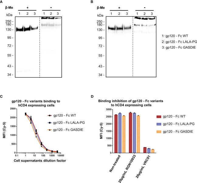
Figure 2.
Expression and functional analysis of gp120 antigens. (A, B) Supernatants of cells transfected with the indicated gp120 – Fc fusion proteins were analyzed by western blots in presence or absence of the reducing agent β-Me. The Fc-fusion proteins were detected by (A) an anti-mouse IgG antibody coupled to HRP and (B) the 2G12 antibody, a human IgG1 to HIV-1 Env followed by an anti-human IgG antibody coupled to HRP. The dotted lines represent the truncation between the blot pictures (C) Four-fold serial dilutions of the cell supernatants containing the gp120 – Fc variants proteins were incubated with hCD4 expressing TZMbl cells. Binding of the Fc-fusion proteins to hCD4 expressing cells were detected via an anti-mouse IgG antibody coupled to Cy-5 and samples were analyzed by flow cytometry. (D) Prior to staining of TZMbl cells, the undiluted cell supernatants containing the Fc-fusion proteins were pre-incubated 1h at 37°C with either 20µg/mL of the CD4bs anti-HIV Env VRC01 antibody, 20µg/mL of the anti-RBD RGN10933 antibody or with DMEM medium (= Non-treated control). TZMbl cells were then stained as described above and binding inhibition was determined by flow cytometry.

