Figure 2.
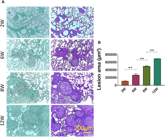
Establishment of granuloma. Groups of four C57BL/6 mice were infected by the IT. route with 1 × 10⁶ P. brasiliensis yeasts contained in 50 µL of PBS. After two (2W), six (6W), eight (8W) and twelve (12W) weeks of infection, the animals were euthanized and the lungs removed, which were stored in 10% formaldehyde at 4 °C. Sections of 5 µm were stained with Hematoxylin-Eosin (H&E; pink) for analysis of lesions and stained with Grocott (green) for fungal evaluation (A). The anatomy of the lesion was analyzed according to size, morphology and presence of fungi. The image and the lesion area calculation were taken using the Leica DM750 microscope and the Leica Application Suite V4.8 software. Statistical analysis was performed using one-way ANOVA. Bars represent means ± standard error of lesion area (µm²) of groups of three mice (B) **p<0.01.

