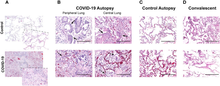
Figure 1.
Changes in air space in central and peripheral lung samples from COVID-19 and control autopsies, and convalescent lung samples. (A) Comparison of lung histologic cross-sections from control and COVID-19 autopsy; 40x magnification with 200x magnification inset. (B–D) Representative photomicrographs of hematoxylin & eosin-stained lung tissue from COVID-19 autopsy (B), control autopsy (C), and convalescent lung tissue (D). 200x magnification (scale bar: 200 microns). Filled arrow: metaplasia of type II pneumocytes; dashed arrow: hyperplasia of type II pneumocytes.

