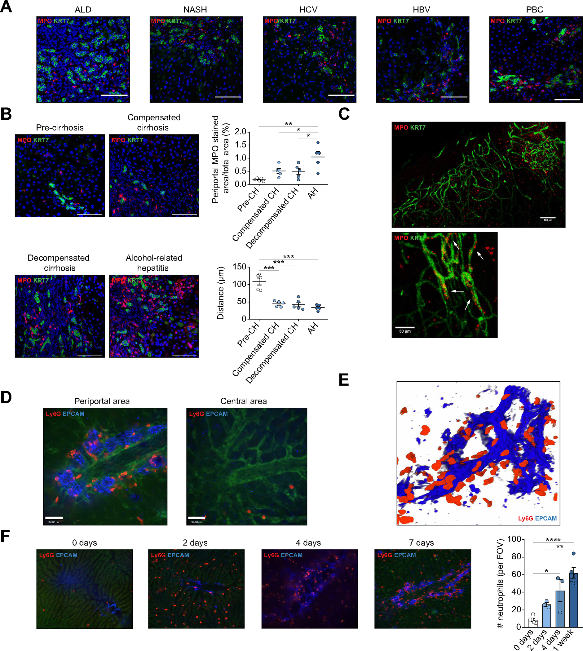
Fig. 1. DRANs are recruited to the biliary epithelium.

(A) Immunofluorescence of MPO and KRT7 in liver sections of patients with ALD, NASH, HCV, HBV and PBC. Scale bar: 100 μm. (B) Immunofluorescence of DRANs (MPO) at biliary epithelium (KRT7) in hepatic biopsies of patients with ALD. Percentage of MPO+ cells at periportal areas and minimum distance of MPO+ cells to KRT7+ cells. Scale bar: 100 μm. (C) Clearing of 3 mm-liver section of a patient with cirrhosis. Arrows show neutrophils (MPO) attached to biliary cells (KRT7). (D) SD-IVM images of periportal and central areas in DDC-treated mice. (E) 3D-reconstruction of neutrophils (Ly6G) recruited to the biliary epithelium (EpCAM) in mice treated with DDC for 1 week. (F) SD-IVM images of DDC-treated mouse showing the progression of neutrophil (Ly6G) recruitment to ductular reaction (EpCAM). All data is presented as mean ± SEM. *p <0.05, **p <0.01, ***p <0.001 as determined by one-way ANOVA with Tukey’s multiple comparison test (B, F). AH, alcohol-related hepatitis; ALD, alcohol-related liver disease; CH, cirrhosis; DDC, 3,5-diethoxycarbonyl-1,4-dihydrocollidine; DRANs, ductular reaction-associated neutrophils; EpCAM, epithelial cell adhesion molecule; FOV, field of view; KRT, cytokeratin; Ly6G, lymphocyte antigen 6 complex locus G6D; MPO, myeloperoxidase; NASH, non-alcoholic steatohepatitis; PBC, primary biliary cholangitis; SD-IVM, spinning-disk confocal intravital microscopy.
