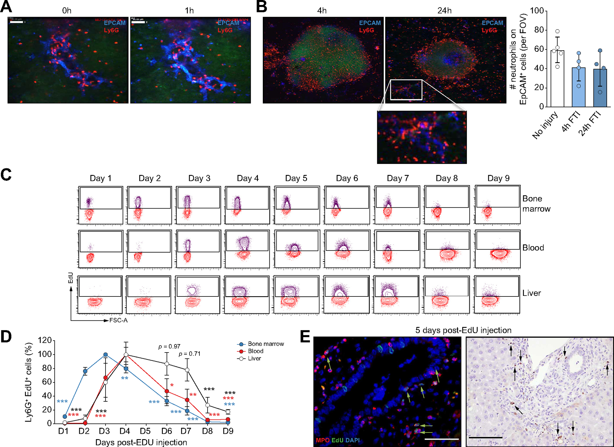
Fig. 2. DRANs are immobilized at the biliary epithelium and remain static.

(A) Time-lapse SD-IVM images showing neutrophil (Ly6G) static behaviour at ductular reaction (EpCAM) sites. (B) SD-IVM images of FTI (Sytox Green) in mice treated with DDC for 1 week. Quantification of neutrophils (Ly6G) retained at EpCAM+ cells per FOV. (C) Flow cytometry plots showing Ly6G+ EdU+ neutrophils. (D) Percentage of Ly6G+ EdU+ neutrophils (n = 3–4 mice per time point). Data presented as mean ± SEM. Each time point was compared by two-way ANOVA with Dunnett’s multiple comparison test vs. day 3 (bone marrow samples) or day 4 (liver and blood samples). *p <0.05, **p <0.01 and ***p <0.001. (E) Immunofluorescence (scale bar: 50 μm) and EdU immunohistochemistry (scale bar: 100 μm) of liver sections of mice fed with DDC after 5 days of EdU injection. EdU, 5-ethynyl-2’-deoxyuridine; FOV, field of view; FTI, focal thermal injury; Ly6G, lymphocyte antigen 6 complex locus G6D; SD-IVM, spinning-disk confocal intravital microscopy.
