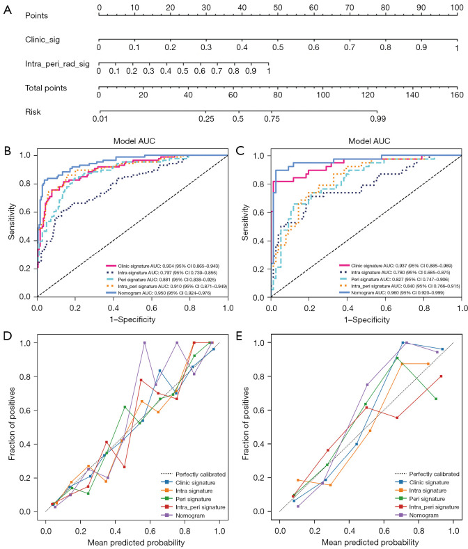
Figure 3.
A radiomics nomogram (A) was developed in which the intratumoral peritumoral ultrasound radiomics were combined with clinical and ultrasound features. The receiver operating characteristics of the radiomics nomogram, intratumoral model (intra signature), and intratumoral combined peritumoral model (intra_peri signature) in the differential diagnosis of breast nodules were compared between the training set (B) and the test set (C). The radiomics nomogram calibration curves for the training set (D) and test set (E). intra, intratumoral features; peri, peritumoral features; AUC, area under the curve; CI, confidence interval.

