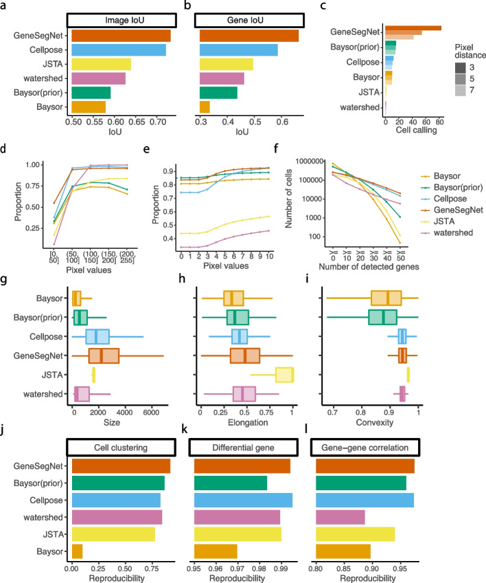
Fig. 7.
Quantitative analysis in NSCLC dataset. a The image IoU scores. b The gene IoU scores. c Cell calling metrics. d Proportion of pixels within cell boundaries (y-axis) for pixels with different levels of pixel values (x-axis). e Proportion of RNA reads within cell boundaries (y-axis) for RNA reads with different pixel values (x-axis). f Number of cells (y-axis) with different numbers of detected genes (x-axis). A gene is detected if it has at least one read in that cell. g–i The distribution of cell size in pixels (g), elongation (h), and convexity (i). j–l Reproducibility of cell clustering (j), differential gene analysis (k), and gene-gene correlations (l) across biological replicates

